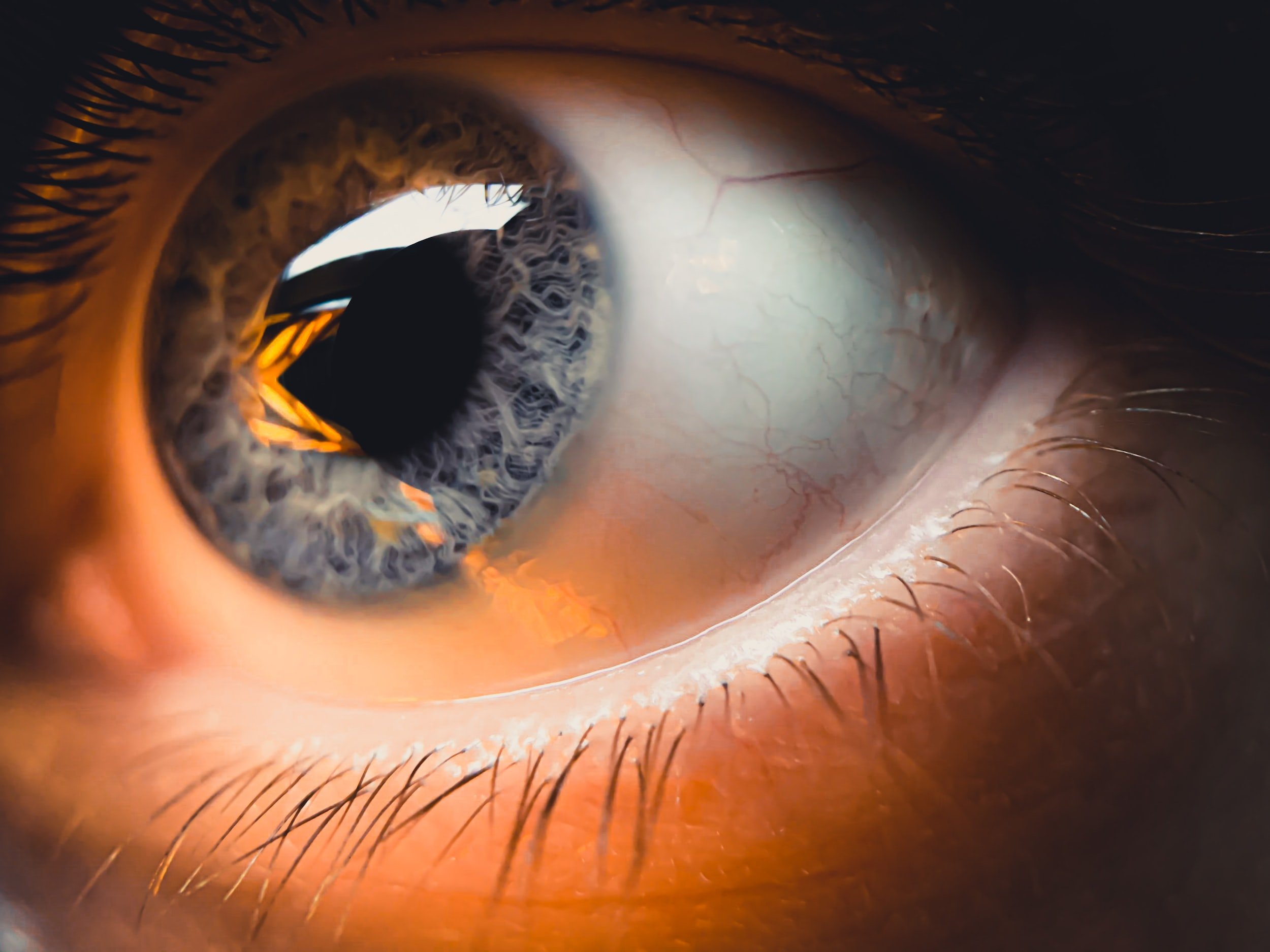
“The eye is the window to the soul, and the cornea is the window to the eye. With corneal donation for transplantation, we are able to provide the gift of sight. ”
Corneal Transplants Philadelphia
Your Sight, Our Passion
Providing the highest quality surgical experience for DMEK, DSEK, PKP, and DALK patients from Montgomery, Bucks, Chester, and Delaware Counties. Easy access from Horsham, Plymouth Meeting, Fort Washington, Collegeville, King of Prussia, Philadelphia, Dresher, Blue Bell, Doylestown, Gladwyne, Bryn Mawr, Villanova, Devon, and Berwyn.

Corneal Transplants at SVI
The cornea is the front layer of the eye. Due to a variety of conditions, the cornea can become cloudy. When this happens, vision becomes blurry and the eye can become painful. Common conditions affecting the cornea include Fuchs Dystrophy, Keratoconus, Pseudophakic Bullous Keratopathy, Corneal Ulcers, and Anterior Basement Membrane Dystrophy. All of these conditions can lead to permanent vision loss if not treated appropriately. While some conditions require replacement of the entire cornea, others require replacement of just the back layer.
Dr. Shafer is fellowship-trained in corneal transplant surgery. During his fellowship, he performed hundreds of transplants, restoring sight to the blind. Since then, he has continued performing advanced corneal transplants for the patients of the Greater Philadelphia area. Dr. Shafer routinely performs both DMEK and DSEK surgery for patients afflicted by Fuchs Dystrophy and Pseudophakic Bullous Keratopathy. Fortunately, the need for full-thickness corneal transplants has diminished in recent years due to advances in corneal transplant technique. Dr. Shafer is up to date on all of the latest approaches and tailors each surgery to the patient’s individual needs.

Frequently Asked Questions about Corneal Transplants
-
The cornea is the very front of the eye. It is the clear structure that people put contact lenses on.
-
Corneal transplant surgery involves removal of one or more layers of the cornea and replacement with donor tissue. There are multiple different types of transplants such as DMEK, DSEK, PKP, DALK, and DSO. They all have their own unique benefits and drawbacks.
-
The cornea is like a sponge. It likes to soak in water. The back layer of the cornea, the endothelium, is responsible for ringing out the sponge to keep it clear and compact. In Fuchs Dystrophy, the cornea fills up with water but the endothelium is unable to pump it out. Therefore, the cornea becomes swollen, cloudy, and painful. The treatment is replacement of the endothelium via either DMEK or DSEK.
-
Pseudophakic Bullous Keratopathy, or PBK, is a condition where the endothelial cells of the cornea die due to complicated surgery. This could be from cataract surgery, glaucoma surgery, dislocated IOL surgery, or even retinal surgery. The treatment involves replacing the back layer of the cornea via either a DMEK or DSEK.
-
Keratoconus is a progressive thinning and bulging of the cornea, typically associated with eye rubbing and genetics. While most patients can have their condition stabilized through the use of scleral lenses and crosslinking, some patients require corneal transplants, either a DALK or PKP.
-
DMEK (Descemet's Membrane Endothelial Keratoplasty) is a type of cornea transplant surgery that replaces the innermost layer of the cornea, called the Descemet's membrane, and the endothelial cells. This is the most anatomical replacement as it involves removal of a sick layer and replacement by that same layer. This is the most commonly performed type of corneal transplant by Dr. Shafer. He has performed hundreds of DMEK surgery and teaches the surgery to surgeons around the country.
-
DSEK, or Descemet Stripping Endothelial Keratoplasty, is similar to DMEK except it involves placement of a thicker piece of tissue. The benefit of DSEK is that it can be utilized in patients with more complex eyes compared to DMEK.
-
DMEK surgery is typically recommended for patients with Fuchs' dystrophy, a genetic disorder that causes the endothelial cells in the cornea to malfunction and die. It is also used for patients with other conditions that affect the endothelial cells, such as pseudophakic bullous keratopathy.
DSEK is preferred for patients who have had prior glaucoma surgery or retina surgery.
-
DMEK surgery can improve vision and reduce the risk of complications compared to traditional corneal transplants. It also has a faster recovery time and a lower rejection rate. DMEK tends to have the fastest and best visual recovery, but can involve some maintenance such as rebubbling procedures.
-
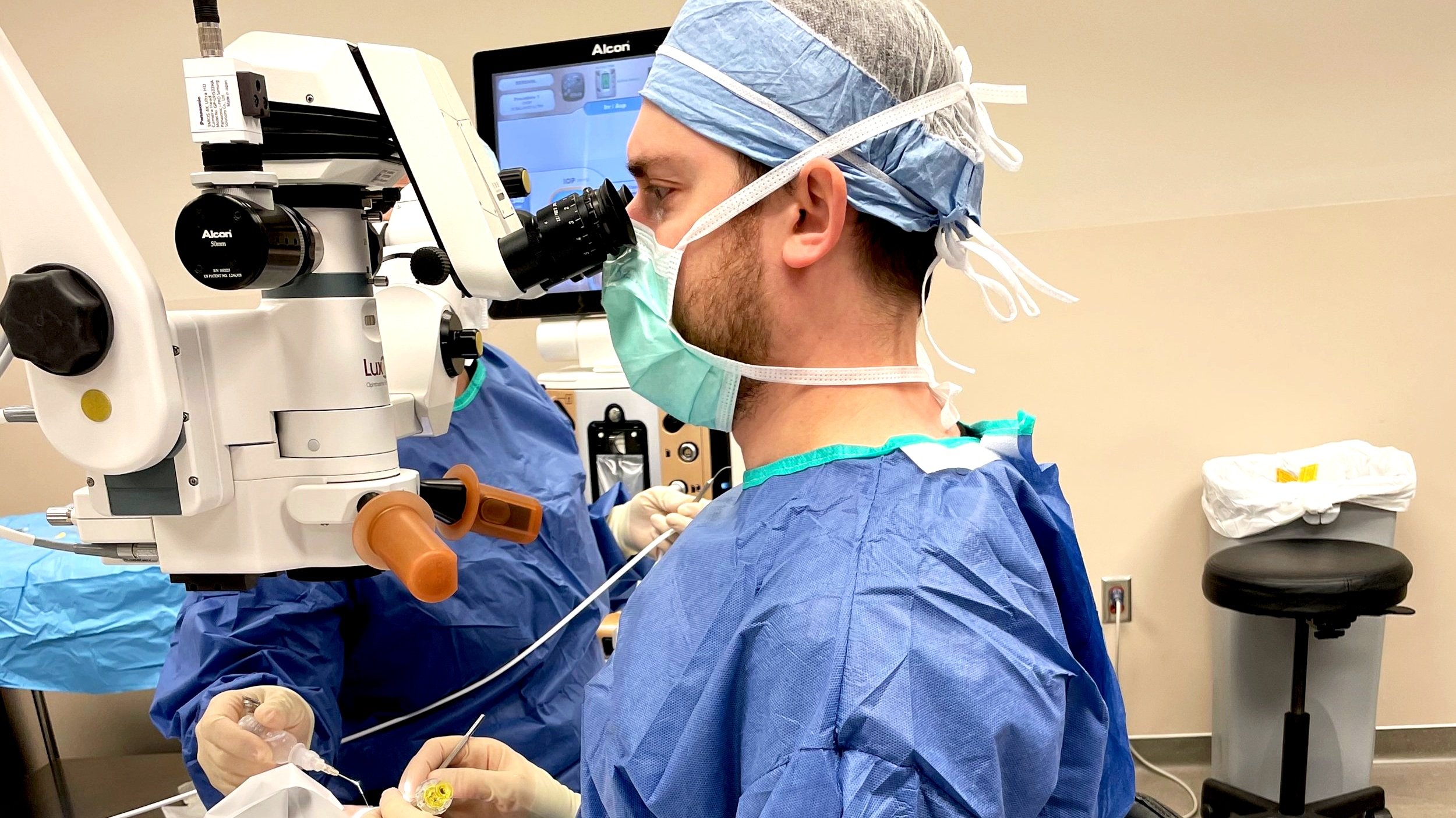
During surgery, Dr. Shafer uses a microscope to carefully remove the damaged inner layer of the cornea and replace it with a healthy donor membrane. After the healthy donor cornea is in position, a large gas bubble will be placed to push the new cornea against the back layer of your cornea. The surgery is typically performed on an outpatient basis and takes under an hour. After surgery, you lay on your back for a full 24 hours. You can be upright for 15 minutes out of every hour. The time on your back is essential in making sure the surgery is successful.
-
When the new cornea is placed inside of the eye, a gas bubble is used to hold it up against the back layer of your cornea. By lying flat on your back, the bubble stays in contact with the transplant and holds it in place while the cells start to “wake up” and attach on their own. After 24 hours, you are allowed to be upright again.
-
For the first 24 hours, you will be lying flat on your back. You are allowed to be upright for 15 minutes out of every hour. Your eye will be uncomfortable. We expect it to be scratchy, blurry, and irritated. In fact, your vision will be extremely blurry until the gas bubble dissolves after a week or so. We do not expect you to have intense eye pressure, nausea, vomiting, or sinus pain. If you have those symptoms, please alert us right away as this could indicate that the pressure in your eye is too high from the gas. We would have to see you right away to take care of this.
-
As with any surgery, there is a risk of complications such as infection, bleeding, or rejection of the donor tissue. The biggest risk is the need for more surgery in the future. There is a 5-10% rate of primary graft failure where the donor tissue never “wakes up.” In this situation, it is necessary to go back to the operating room to repeat the surgery with a different donor. Although this leads to prolonged recovery, it is not typically long-term visually significant. Other risks include glaucoma, cataracts, and vision loss. These are rare and will be monitored closely in the post-operative period.
-
Recovery time varies from person to person, but most patients can return to their normal activities within a few days of the surgery. It may take several weeks for the vision to fully recover. If the vision is not clearing completely in the first couple of weeks, it may be necessary to place more air inside of the eye to help facilitate attachment of the graft. This occurs in the office and needs to be done around 50% of the time at the one-week mark.
-
At the one-week mark after surgery, we hope that the donor cornea has fully attached to your natural cornea. Once the cornea is fully attached, the vision starts to clear quickly. However, 50% of the time this does not happen right away. To get the cornea to “wake up” more quickly, it is often necessary to place more air into the eye. This is done in the office with numbing eye drops to ensure that you are comfortable. After the rebubble, we will check your pressure and then send you home. Although not as essential, the more time you lay on your back for the first 24 hours after a rebubble the better.
-
Approximately one week prior to surgery, Dr. Shafer will perform what is called a Laser Peripheral Iridotomy (LPI) in the office. This is a quick laser procedure that creates a small hole in the iris. This helps prevent a large spike in pressure when the gas bubble is placed in the eye. After the laser, you will start your steroid eye drops that you will be on for the full year.
-
For the first 24 hours, you will have to lay flat on your back. For the first week after surgery, you are not allowed to lift anything greater than 10 pounds, bend at the waist, or participate in any vigorous activity. You are not allowed to fly until the gas bubble in your eye has completely disappeared, which usually takes 1-2 weeks.
-
After surgery, you will require an antibiotic eye drop for around one week and a steroid eye drop to prevent rejection for the first full year after surgery. The drop will typically be used four times per day for the first three months, and then will be tapered down from there. Some patients require steroid eye drops for a lifetime to prevent rejection.
-
While DMEK surgery can significantly improve vision, it may not be perfect. Most patients will need to wear glasses or contact lenses after the surgery.
-
Most insurance plans cover DMEK surgery, but it is important to check with your insurance provider to confirm coverage.
-
PKP stands for penetrating keratoplasty. This is the formal term for a full-thickness corneal transplant. Removal of the entire cornea and replacement with a donor cornea is a serious surgery that requires 16 or more stitches and a year of recovery. After surgery, patients may still require a specialty contact lens for best vision. There is a lifelong risk of rejection and dehiscence, or wound leak.
-
DALK, or Deep Anterior Lamellar Keratoplasty, involves the removal of the outer layers of the cornea without making a full-thickness incision as with PKP. This is a condition reserved for keratoconus. Not all surgeries are amenable to DALK and often result in a PKP. There are some theoretical benefits to DALK over PKP such as lower risk of rejection and lower risk of wound leak.
What Are You Waiting For?
Trust the Best With Your Cornea
Schedule your free consultation now
Specialty Surgery at SVI
Refractive
Surgery
Correct your vision with our cutting-edge refractive surgery options, including LASIK, PRK, RLE, and ICL. Dr. Shafer utilizes state-of-the-art equipment to help you achieve clear vision, reducing or eliminating the need for glasses or contacts.


